Nils Jacobi / Shutterstock.com
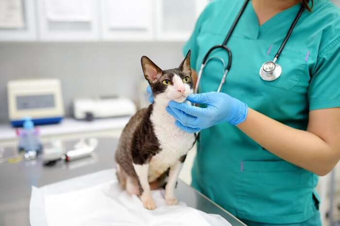
Liver flukes are a type of worm-like parasite that can cause liver and bile duct infections in many animals, including cats. In this article, you’ll learn about the most common types of flukes that can affect cats, the regions where they occur most often, and how they’re diagnosed and treated.
Quick Overview: Liver Flukes in Cats
 Other Names: “lizard poisoning” (only for Platynosomum fastosum)
 Common Symptoms: Mild signs ranging from weight loss to intermittent digestive upset signs. More severe signs of liver disease may include jaundice, abdominal distention, and more severe signs of digestive upset.
 Requires Ongoing Medication: No
 Vaccine Available: No
 Treatment Options: Praziquantel is used most often for Platynosomum Fenbendazole may be appropriate for some other types of liver fluke infections.
 Home Treatment: Signs of jaundice (yellow skin, eyes, gums) and anorexia (no appetite) should always be considered emergencies in cats, regardless of cause. Liver flukes are an uncommon cause of these signs in cats. The region where you live can factor greatly into whether a fluke infection is likely or not. Highest risk cats are those hunting lizards, frogs, and fish. Keeping a cat indoors can greatly reduce risk in higher risk regions.
What’s a Liver Fluke?

What a liver fluke looks like under a microscope. D. Kucharski K. Kucharska / Shutterstock.com
Unlike how we normally think of the word “fluke”, a fluke in the parasite world is not a surprising stroke of luck. Just the opposite, flukes are a type of flatworm parasite that can cause infections. Most often this is in either the intestines or the liver and bile ducts. Flukes are also called trematodes.
Liver flukes are those that specifically migrate to the liver and bile ducts of affected animals, where they live, causing progressive inflammation and damage over time.
There are several different types of liver flukes that occur in cats throughout the world but only a couple are seen in the United States. In this article, we will mention all of them but focus primarily on the one of chief importance in the US, Platynosomum fastosum, the cause of “lizard poisoning” in cats.
Types of Liver Flukes that Affect Cats
1. Platynosomum Fastosum
This liver fluke is the one of chief importance in the US. Because ingesting infected lizards is one route that cats can acquire infection, the illness it causes is also known as “lizard poisoning”.
fastosum occurs primarily in Florida, other warm, topic areas of the southeast US, and Hawaii. You may also see it referred to as P. concinnum. They are essentially the same liver fluke. Another called P. illiciens that occurs in Thailand is considered the same parasite, but is a variation occurring more in Southeast Asia.
2. Opisthorchis Felinus
Opisthorchis felinus is a liver fluke found primarily in Italy, Eastern Europe, Russia, and parts of Asia. In these regions, it can be a significant cause of bile duct inflammation and infection (cholangitis) in cats who catch and ingest raw fish. The same route of infection can also occur in people, causing the condition opisthorchiasis.
3. Clonorchis Sinensis
Clonorchis sinensis, known as the oriental liver fluke that infects people, has also been found in the bile and pancreatic ducts of dogs and cats across parts of Asia.
4. Metorchis Orientalis/Conjunctus
Metorchis orientalis/conjunctus are very small liver flukes that have been found in the bile ducts and gallbladder of carnivores, including dogs and cats. They have been found in North America, but are not considered significant in the US. They also occur in Europe, Russia, and China.
Eurytrema Procyonis
Eurytrema procyonis is a liver fluke that mainly affects raccoons and foxes in the Eastern US. It has been found sometimes in the pancreatic duct, bile duct, and gallbladder of domestic cats.
Causes of Liver Flukes in Cats

Cats who hunt small anole lizards like this one, a common finding in Florida and similar regions, are at higher risk for liver fluke related disease. Chris Mercer / Shutterstock.com
Cats get infected by liver flukes through ingesting a smaller animal that has the infective stage (called metacercariae) in its body. These smaller animals are called intermediate hosts. Flukes require 1-2 intermediate hosts as part of their life cycle.
Snails are common first intermediate hosts. They ingest fluke eggs passed in stool by infected mammals. The snails are then eaten. Cats could get infected at this stage, but snails aren’t super appetizing for them. A second intermediate host more often ingests the snails and that second intermediate host is then eaten by the affected cat.
Second intermediate hosts can include fish, amphibians, and reptiles. Fish are the main intermediate host for Opisthorchis felinus. For Platynosomum fastosum, cats acquire the parasite through hunting and eating frogs and lizards.
Symptoms of Liver Flukes in Cats
When cats eat a lizard or frog acting as an intermediate host, the infective stage is a type of cyst called metacercariae. After ingestion, these cysts will “hatch”, allowing the baby flukes to emerge. For liver flukes (as opposed to intestinal flukes) the juveniles will find their way to the bile duct (which opens into the small intestine) and migrate through those ducts to the gall bladder.
In cats, liver flukes may also migrate up the pancreatic duct, owing to the pancreatic duct and bile duct sharing a common opening into the small intestine.
Liver flukes may make their way to the gallbladder. They also migrate from the main larger common bile duct to any of numerous branches of bile ducts spread throughout the liver.
It takes about 8-12 weeks for the younger flukes to develop into mature adults.
The flukes’ location contributes to signs of disease seen. Many cats may have no apparent symptoms of illness at first. But over time, the presence of the flukes cause blockages in the bile ducts (and sometimes pancreatic ducts), causing obstruction of bile flow, tissue damage and inflammation. These changes can also lead to secondary bacterial infection as well.
Signs may occur as early as 7 days after infection but may take up to 4 months.
While cats in earlier stages of infection or with a light burden of flukes may show no signs of illness or mild intermittent signs of illness:
- Mild weight loss or inability to gain weight
- Fluctuations in appetite
- Intermittent diarrhea
- Poor haircoat
- Intermittent lethargy
- Intermittent low grade fever
Complications Occurring in Cats with Liver Flukes

Jaundice, or a yellow appearance to the skin, gums, and whites of the eyes, is a sign of liver and bile duct disease, which liver flukes can cause. Todorean-Gabriel / Shutterstock.com
The degree of disease depends on how many liver flukes are present and how long a cat has been infected. More severe infections in cats lead to signs of liver and gallbladder/bile duct disease, which is called hepatobiliary disease or cholangiohepatitis. By 4-6 months after infection, flukes have caused significant tissue changes in the liver and bile ducts, leading to more noticeable signs of illness.
In cats where these signs are caused by Platynosomum, these signs of illness are collectively called “lizard poisoning”:
- Lack of appetite (anorexia)
- Lethargy
- Vomiting
- Diarrhea
- Jaundice (yellow tinge to gums, whites of eyes, and skin)
- Abdominal distension
Cats that develop signs of illness to this extent will die from the disease if it goes untreated.
It is important to remember that these signs of illness are not exclusive to lizard poisoning but can be seen with any cause of liver and/or gallbladder disease in cats.
Diagnosis of Liver Flukes in Cats

Special fecal testing methods are needed to see liver fluke eggs in a stool sample. Peerachart / Shutterstock.com
Diagnosing liver flukes can be very challenging. Knowing the region and likelihood of exposure for a cat is important. But there are diagnostics used to further narrow down the possibility of liver flukes being involved in a cat’s illness.
1. Fecal Testing
Cats with high parasite loads are likely to have eggs found in their feces, but it will still take at least 12 weeks for eggs to start showing up in stool samples after infection.
Flukes require specific methods of fecal detection with the following techniques:
- Fecal flotation using Sheather’s sugar solution
- Fecal sedimentation
These methods are not routinely used in veterinary practice for a majority of parasites that can be identified with fecal exams (because these methods don’t work well for identifying far more common parasites). Thus flukes can be easily missed using routine fecal exam techniques, especially for cats with very light burdens of flukes.
Parasites like flukes shed eggs intermittently, which can lead to false negative test results if the parasite simply isn’t producing eggs at the time of the fecal exam. Multiple fecal exams using the right techniques may be needed.
In cases where flukes are causing bile duct obstruction (which can occur with severe disease), egg shedding may not occur at all.
PCR (DNA) testing is becoming more common to test for fecal parasites. To my knowledge there is no such testing available for cat liver flukes. Liver flukes can be identified through PCR testing. However, because liver flukes are not intestinal parasites living in the digestive tract, PCR testing on stool may not be accurate.
2. Blood Testing
There is no specific blood test for liver flukes in cats. We can see other blood work changes that occur because of the disease the flukes are causing in affected cats.
ALT: This is a liver cell leakage enzyme that increases with liver inflammation when the cells are under stress. This can occur with any kind of hepatic (liver) disease. Another less specific value called AST is also often elevated with ALT.
ALP: Is a less specific value on blood work but can be seen with hepatobiliary disease. This usually only starts to elevate with more severe bile duct disease.
Total Bilirubin (TBili): Without getting technical, total bilirubin is a value that will increase with severe gallbladder or bile duct disease, especially blockage or obstruction. Even a mild increase in total bilirubin is considered highly abnormal and concerning in cats.
Eosinophils: Eosinophils are a type of white blood cell that will see increases with parasitic diseases. One study looking at liver flukes in cats found that eosinophil counts were increased in all cats with fluke infection but peak levels took 4-5 months, making this a less sensitive indicator for early disease.
3. Imaging
X-ray is not helpful for diagnosing liver fluke infection. But ultrasound can be very helpful, especially for cats with more advanced disease where changes in the liver and bile ducts may be visible.
Ultrasound is a routine diagnostic imaging test used to evaluate cats with hepatobiliary disease. In the case of liver fluke infection, changes like bile duct and gallbladder enlargement may be seen. While challenging to find, the presence of a high burden of flukes may actually be visible to a degree within distended bile ducts. Regional lymph nodes may also be enlarged and detectable only through ultrasound.
Because fecal testing may be less reliable, sampling of bile (which could be done through ultrasound) has been suggested as a possible way to diagnose flukes.
Treatments for Liver Flukes in Cats
Because testing for liver flukes can be very challenging, treating cats perceived to be at risk may be the best course of action in some cases.
For Platynosomum liver fluke infections, praziquantel has demonstrated the best success. It is not approved for this use, but a 3-5 day course repeated after 12 weeks has been suggested.
For other types of fluke infections, fenbendazole has also been described as a treatment option.
For very heavy liver fluke infections causing bile duct obstruction, surgery may be required.
Any cat that is very ill from hepatobiliary disease often requires additional supportive care including:
- Intravenous or subcutaneous fluids
- Nutritional support (even feeding tubes for cats that are not eating)
- Antibiotics
- Steroids
- Appetite stimulants
- Medication for vomiting and/or diarrhea.
Cat Care Tips
- Liver flukes in cats are not a common cause of hepatobiliary disease in cats except in regions where these parasites are found (southeast US and tropic areas)
- If you have a rescue cat that develops signs of hepatobiliary disease within only a few weeks or months, always check if they may have originated from a higher risk region.
- Non-specific signs of illness can occur commonly in cats. But if these signs occur off and on over and over (even if your cat appears normal in between) it’s a good idea to get a veterinary exam.
- Any time a cat stops eating for more than 24 hours, you should always consider this an urgent need for a veterinary exam
- Any signs of jaundice (yellow tinge to skin, gums, and whites of eyes) are associated with severe liver or bile duct disease and should be considered an emergency.
Prevention of Liver Flukes in Cats

Cats who hunt lizards, frogs, and fish are at higher risk of liver fluke infection. Kristi Blokhin / Shutterstock.com
Cats can only acquire liver flukes through ingestion of the intermediate host (lizards or frogs for Platynosomum and fish for Opisthorchis). Therefore, cats who are hunting these types of animals are at highest risk.
Keeping cats in these more tropic regions indoors can virtually eliminate the risk.
In cases where the risk is high but hunting cannot be eliminated from a cat’s routine, screening fecal samples with the appropriate techniques could be considered every 12-16 weeks.
If the risk is high enough, empirical treatment with a praziquantel product every 12 weeks may be a consideration.
If you live in a region where liver flukes may present a higher risk of illness, make sure to discuss options with your local veterinarian.
-
Opisthorcus felinus. (2024, December 14). Wikipedia.
-
A Center, S. (2023, August). Hepatobiliary Fluke Infection in Small Animals. Merck Veterinary Manual, Professional Version.
-
S Peregrine, A. (2023, July). Flukes in Small Animals. Merck Veterinary Manual, Professional Version.
-
Companion Animal Parasite Council (CAPC). (2019, March 13). Platynosomum fastosum.
-
Basu, A. K., & Charles, R. A. (2014). A review of the cat liver fluke Platynosomum fastosum Kossack, 1910 (Trematoda: Dicrocoeliidae). Veterinary Parasitology, 200(1–2), 1–7. https://doi.org/10.1016/j.vetpar.2013.12.016
-
Rothuizen, J. & World Small Animal Veterinary Association World Congress. (2006). Cholangitis in Cats: A Review. Veterinary Information Network.
-
CDC. (n.d.). Opisthorciasis. Centers for Disease Control (CDC).
-
Chantawong, P., Potiwong, J., Choochote, N., Piyarungsri, K., Kunkaew, C., Tangtrongsup, S., & Tiwananthagorn, S. (2024). Occurrence and Risk Factors Associated with Platynosomum illiciens Infection in Cats with Elevated Liver Enzymes. Animals, 14(7), 1065. https://doi.org/10.3390/ani14071065
-
Fecal Sedimentation: Purpose. (n.d.). The RVC/FAO Guide to Veterinary Diagnostic Parasitology.